Actinic keratosis: what it looks like, whether it is dangerous and how it is treated
A spot on the forehead, nose or crown of the head that feels rough to the touch, occasionally peels, and then returns to the same spot after a few weeks. It is not painful and does not grow rapidly, so for months it is attributed to dry skin. Very often it is actinic keratosis, a skin lesion prepared by the sun over years that, when recognised in time, is resolved simply.
Actinic keratosis in brief
- Actinic keratosis is a rough, scaly patch on the skin that has been exposed to the sun for years. It is also called solar or sun keratosis.
- It most commonly occurs on the face, ears, scalp with thinning hair, lower lip, backs of the hands and forearms.
- It is considered a pre-cancerous stage of cutaneous squamous cell carcinoma. A single lesion rarely develops into cancer, but it is impossible to predict which one will, so treatment is generally recommended.
- One or more lesions are most commonly treated with liquid nitrogen. When there are several in the same area, the whole region is treated with a prescription cream or photodynamic therapy.
- A change that thickens, becomes painful, bleeds or grows rapidly requires examination without delay.
What is actinic keratosis
Ultraviolet radiation accumulates in the skin throughout a person's life. After enough years, the cells of the superficial layer of skin in the most exposed areas begin to change, forming a localised, rough patch. This is actinic keratosis.
Different names are found in literature and findings: solar keratosis, sun-induced keratosis, senile keratosis, and in Croatian texts actinic keratosis. All of them refer to the same lesion.
What does actinic keratosis look like
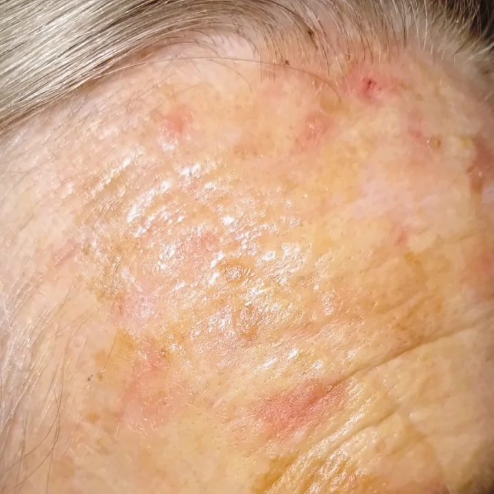
Most commonly it is a pinkish, reddish or brownish patch, ranging in size from a millimetre or two to a centimetre and more, with a dry scale that adheres firmly to the skin. Many are easier to feel than to see: under the finger they feel like fine sandpaper. Some are flat, some thickened, and some have a hard outgrowth resembling a small horn. They may be sensitive to touch, but most cause no discomfort.
It is rarely just one. Whoever finds one usually finds several more in the same region.
On the face, nose and ears
The forehead, temples, cheeks, nose and upper lip are the places where people most often notice them themselves, usually as redness that flakes and does not go away after moisturiser. On the edges of the ears they are easily missed, as that area is rarely looked at in the mirror.
On the crown
In men with thinning hair, the crown has been unprotected for years. Changes often appear there in clusters, and the skin between them is thin, mottled and finely textured.
On the bottom lip
This change on the lower lip is called actinic cheilitis. The lip is permanently dry, peels and cracks, and the border between the red part of the lip and the skin becomes blurred.
On the backs of the hands and forearms
The lesions on the hands and forearms are usually thicker and harder than on the face, and they respond more slowly to treatment.
Who gets actinic keratoses
Most commonly, fair-skinned people who burn easily, those who have spent years working or spending their leisure time outdoors, and people with a weakened immune system, for example after an organ transplant. Among fair-skinned people older than 50, it occurs in more than a third of cases, so it ranks among the most common reasons for visiting a dermatologist.
Actinic or seborrhoeic keratosis
Both are called keratosis, but they differ in what matters most.
| Actinic keratosis | Seborrhoeic keratosis | |
|---|---|---|
| Appearance | Flat or slightly thickened, rough, with a dry scale | Clearly raised, waxy, as if stuck onto the skin |
| Colour | Pink, reddish, skin-coloured, more rarely brown | Light brown to almost black |
| Where does it occur | Only in places exposed to the sun | Anywhere except the palms and soles |
| Attitude towards skin cancer | Pre-invasive stage of squamous cell carcinoma | Benign, does not turn into cancer |
| Reason for treatment | Medical | Most commonly for aesthetic reasons or because it's bothersome |
At first glance, they are not always easy to distinguish, especially when actinic keratosis is pigmented. We write about seborrhoeic keratoses and their removal on the page Removal of keratoses.
Is keratosis malignant
The answer depends on which keratosis is being referred to. Seborrhoeic keratosis is benign. Actinic keratosis is not cancer, but it is a lesion from which squamous cell skin carcinoma can develop. It is not linked to melanoma in that way.
For a single lesion in one year the risk is small: recent analyses estimate it at less than 0.1 per cent, and at around 0.5 per cent in people who have already had skin cancer. Some lesions even resolve on their own. The picture changes when there are many of them and they persist for years: in individuals with more than ten actinic keratoses, the probability of developing squamous cell carcinoma at some point is estimated at 10 to 15 per cent.
Two things explain why dermatologists still recommend treatment. It is impossible to predict from appearance which lesion will progress. In addition, visible keratoses are only the most noticeable part of skin that is entirely sun-damaged, so cancer can also develop in an area that appears healthy. Therefore, the focus is not on just a single spot, but on the entire region, and during the examination, the rest of the skin as well.
Signs that mean an examination should not be delayed
- the lesion has thickened or is hard to the touch
- it became painful or sensitive
- there is a small sore on it that does not heal or bleeds occasionally
- is increasing rapidly
- has darkened or changed colour
- she returned to the same place after treatment
None of these signs on their own means that it is cancer, but each one is sufficient reason for a sample to be taken for analysis.
How is the diagnosis made?
In most cases, an examination is sufficient. The dermatologist looks at and feels the lesion, and then examines it with a dermatoscope, a device that magnifies and illuminates structures invisible to the naked eye. More about that on the page Dermoscopic examination.
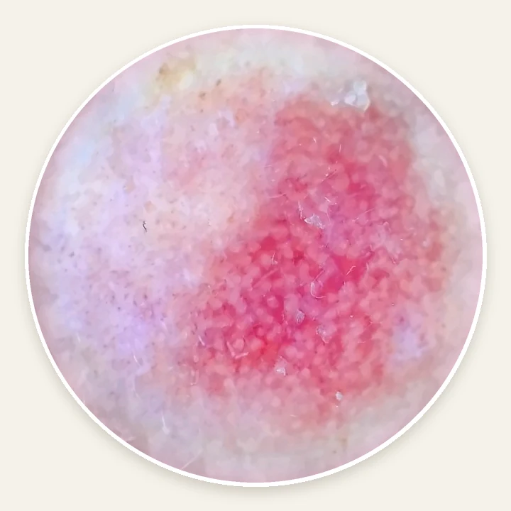
A biopsy is not performed routinely. It is taken when a lesion appears suspicious, when it has any of the aforementioned signs or when it does not respond to treatment. In individuals with a weakened immune system, a biopsy is decided upon earlier. We describe the procedure on the page Skin biopsy.
How is actinic keratosis treated
The choice depends on the number of lesions, their thickness and location, as well as how much time and patience the patient has for the therapy. Therefore, the decision is made through a discussion, not by following a set formula.
One or several changes
Standard treatment is cryotherapy with liquid nitrogen. Freezing lasts a few seconds, after which redness appears, sometimes a blister, followed by a scab that falls off. On the face, the skin heals in 5 to 10 days, on the hands in 3 to 4 weeks, and on the shins in six weeks or longer. Brief freezing of a superficial lesion usually leaves no trace, while a longer one may leave a lighter patch.
Thickened lesions and those with a horny outgrowth are removed under local anaesthetic, and the tissue is sent for histopathological analysis. An ablative laser is another option, although it has not shown any advantage over cryotherapy.
Multiple changes to the same region
When there are five, ten or more of them on the forehead or crown, freezing them one by one does not solve the problem, because the skin between them is also damaged. In that case, the entire region is treated. The good side of this approach is that the therapy also acts on lesions that are not yet visible.
| Therapy | How it is carried out | What to expect |
|---|---|---|
| Fluorouracil 5% cream | Once or twice a day, usually for 3 to 4 weeks | The best long-term results in comparisons, alongside a pronounced skin reaction |
| Imiquimod 5% cream | Three times a week for 4 weeks, followed by a break and a follow-up; if necessary, another cycle | Redness, scabs and scaling; intended for blemishes on the face and scalp |
| Diclofenac 3% gel | Twice daily, 60 to 90 days | The mildest reaction, but also the weaker performance |
| Tirbanibulin 1% ointment | Once daily, for 5 days, on a small area of the face or scalp | Short-term therapy, reaction mostly mild and short-lived |
| Photodynamic therapy | A cream that makes cells sensitive to light, followed by illumination with a lamp or exposure to daylight | It is carried out in a facility; the daylight variant is almost painless. |
All the creams from the table are available on prescription, and which one the dermatologist will suggest also depends on what is currently available in Serbia.
In a major trial involving 624 patients with head lesions, four approaches were compared. One year after the end of treatment, success was maintained in 74.7 per cent of those treated with fluorouracil, 53.9 per cent of those treated with imiquimod, and 37.7 per cent of those treated with photodynamic therapy. This difference is one of the reasons why fluorouracil is often chosen first, but it is not the only criterion: for someone else, it may be more important that the treatment is shorter or that the reaction is milder.
What the skin looks like during cream treatment
This is the part that is most frequently asked about. With fluorouracil and imiquimod, the reaction is part of the treatment: usually in the second week the skin turns red, then sores and crusts appear, followed by healing. Areas where nothing was visible also react, which shows how many lesions there were beneath the surface. During this period the skin looks worse than before the therapy, so the start is planned so that it does not fall before an important event or trip.
Sunlight intensifies the reaction, so the treated area is protected while the treatment is ongoing. Fluorouracil is not used during pregnancy and breastfeeding. If the reaction becomes stronger than was agreed upon at the check-up, the treatment is paused or adjusted in consultation with a dermatologist.
Treatment of actinic keratosis at Imedic
The examination begins with a consultation and an examination of all sun-exposed skin, using a dermatoscope. Individual lesions are most often removed immediately with liquid nitrogen, during the same appointment. Thickened and suspicious lesions are removed under local anaesthetic and sent for histopathological analysis. When there are multiple lesions, the dermatologist prescribes topical treatment, explains what the skin will look like from week to week and schedules a follow-up appointment.
| Service | Price |
|---|---|
| Dermatologist appointment | 5,000 RSD |
| Follow-up appointment | 3.000 RSD |
| Cryotherapy with liquid nitrogen (consultation charged separately) | 2,000 to 12,000 RSD, according to the number of changes |
| Skin biopsy with histopathological analysis | 12.000 RSD |
All prices are in dermatology price list.
Sun protection and tracking
Sun protection here is not just prevention, but also part of the treatment. In a randomised trial, people who regularly used sunscreen over the course of one summer developed fewer new lesions, and existing ones resolved more often than in those who applied a cream without a protective filter.
- sun cream with an SPF of 30 or higher, preferably 50+, every day on your face, ears, neck and hands
- lip balm with SPF
- comb-over
- hunger in the middle of the day and without the sunbed
The treatment outcome is assessed about three months after therapy ends. After that, the schedule of check-ups depends on the number of lesions, whether there were any previous skin tumours, and the state of the immune system. People with a weakened immune system come more frequently, every three to six months. Between check-ups, it is useful to examine the face, ears, scalp and hands once a month, both visually and by touch.
Have a question?
Frequently asked questions about actinic keratosis
Which cream helps with actinic keratosis?
Prescription medicines help: fluorouracil, imiquimod, 3 per cent diclofenac gel and tirbanibulin. Diclofenac gels used for joint pain are not the same preparation and are not intended for this lesion. Urea moisturisers soften the scale and can prepare the skin for treatment, but they do not cure the keratosis itself.
Can actinic keratosis go away on its own?
Agreed. In follow-ups over the course of a year, between 15 and 63 percent of individual changes regressed, but a significant portion of them returned later. Since it is not known which ones will disappear and which ones will progress, waiting only makes sense with an agreement and monitoring by a dermatologist.
Is he returning after medical treatment?
It can return, after several months or years, and new ones may also appear in the same region. That does not mean the treatment was wrong, but rather that the skin as a whole has been damaged by the sun. The same therapy can be repeated or another one chosen.
Is actinic keratosis cancer?
No. It is a lesion from which cancer can develop, specifically planocellular carcinoma. In the case of a single lesion, this rarely happens.
Can I remove the plaster myself?
The scale can be removed, but it returns because the change is not in the scale itself, but in the skin layer beneath it. Removing it with a fingernail or wart treatments only results in a sore in an area that will subsequently be more difficult to assess during an examination.
Can it be treated in the summer?
Cryotherapy is carried out all year round. Cream therapy is more often planned for autumn and winter, as the sun intensifies the skin reaction. The exception is daylight photodynamic therapy, which requires a warmer and brighter time of year.
Does vitamin B3 help?
Nicotinamide, a form of vitamin B3, has been tested in high-risk individuals who have already had skin tumours. On 500 mg twice a day, they had 23 per cent fewer new skin cancers and fewer actinic keratoses than the placebo group, and the effect lasted only while it was being taken. In organ transplant recipients, the benefit was not confirmed. It is not a substitute for treatment and sun protection, and whether it makes sense in a specific case is a topic for a check-up.
Should I go to the doctor?
Dermatologist. Actinic keratosis is diagnosed, treated and monitored in a dermatology clinic.
Book a consultation
If you have a rough patch that flakes and keeps coming back, or a lesion that has recently changed, the quickest route to an answer is dermatological examination. Examinations are carried out by Dr Sanja Kekić and Dr Marina Ćirović, specialist dermatovenereologists.
You can send a photograph of the change to Viber or WhatsApp, and we will tell you which appointment to book.
Telephone, Viber and WhatsApp: 064 237 0707
Address: Kneza Miloša 23, 2nd floor, Belgrade
Opening hours: Monday to Saturday 9am to 9pm, Sunday 12pm to 9pm

Author: Dr Miloš Živković
Doctor in specialist studies of aesthetic, plastic and reconstructive surgery at the Faculty of Medicine, University of Belgrade; in practice she applies radiowave surgery and fractional CO2 laser.
Updated:

Expert review: Dr Sanja Kekić
Specialist in dermatovenerology; graduated from the Faculty of Medicine, University of Belgrade in 2016.
Updated:
Last checked: 19 November 2026.
Expert sources
- Kandolf L. et al. European consensus-based interdisciplinary guideline for diagnosis, treatment and prevention of actinic keratoses, epithelial UV-induced dysplasia and field cancerization. J Eur Acad Dermatol Venereol. 2024. https://pubmed.ncbi.nlm.nih.gov/38451047/
- Jansen MHE, et al. Randomised Trial of Four Treatment Approaches for Actinic Keratosis. N Engl J Med. 2019. https://www.nejm.org/doi/full/10.1056/NEJMoa1811850
- Thompson SC. i sar. Reduction of Solar Keratoses by Regular Sunscreen Use. N Engl J Med. 1993. https://www.nejm.org/doi/full/10.1056/NEJM199310143291602
- Chen AC. i sar. A Phase 3 Randomised Trial of Nicotinamide for Skin-Cancer Chemoprevention. N Engl J Med. 2015. https://www.nejm.org/doi/full/10.1056/NEJMoa1506197
- Allen NC. i sar. Nicotinamide for Skin-Cancer Chemoprevention in Transplant Recipients. N Engl J Med. 2023. https://www.nejm.org/doi/full/10.1056/NEJMoa2203086
- DermNet. Actinic keratosis. https://dermnetnz.org/topics/actinic-keratosis
- DermNet. Seborrhoeic keratosis. https://dermnetnz.org/topics/seborrhoeic-keratosis
- PIL. Summary of Product Characteristics: Efudix 5% cream (fluorouracil). https://www.alims.gov.rs/doc_file/lekovi/smpc/515-01-01362-19-001.pdf
- ALIMS. Summary of Product Characteristics: Aldara 5% cream (imiquimod). https://www.alims.gov.rs/doc_file/lekovi/smpc/515-01-02874-21-001.pdf


