Radiofrequency Mole Removal in Belgrade
Radiofrequency mole removal can be an option for selected lesions that a dermatologist, following a clinical and dermoscopic examination, assesses as suitable for this method. It is most commonly considered when a lesion is bothersome, catches on clothing, or is a cosmetic concern. The choice of procedure, anaesthesia, and post-intervention care is tailored to the specific lesion and the patient.
Radiofrequency mole removal – at a glance
| When it is done | For selected lesions that a dermatologist assesses as suitable for this method following a clinical and dermoscopic examination |
|---|---|
| Anaesthesia | Local anaesthetic or anaesthetic cream |
| Duration | The removal itself takes just a few minutes; the whole appointment, including the consultation and anaesthetic, takes longer |
| Stitches | Not required |
| Analysis | The removed tissue is sent for histopathological analysis |
| Procedure performed by | Dermatovenereology specialist |
What are moles?
Moles are mostly harmless skin lesions caused by the accumulation of melanocytes, pigment-producing cells. Most people have between 10 and 50 moles; they can be flat or raised, light to dark brown, black, pink or skin-coloured. Some are barely noticeable, while others are an aesthetic nuisance or located in places where they are easily irritated. More about the types of moles, monitoring them and choosing a removal method can be found in our article Moles: Check-ups, Dermoscopy & Removal – Dr Ćirović | Imedic.
Who is a candidate for radiofrequency removal?
The radiofrequency method is considered for moles that the dermatologist, after an examination, assesses as harmless and suitable for this procedure. Most commonly, these are raised moles, pedunculated moles, and smaller lesions that cause functional or aesthetic issues – they catch on clothing or jewellery, get irritated during shaving, or the patient dislikes their appearance.
Not every mole is suitable for this method. The choice depends on the type of lesion, its depth, size and location, as well as the doctor's assessment. Before a decision is made, a dermoscopic mole checkis performed, and when there is any doubt or the lesion needs to be removed in its entirety for analysis, priority is given to surgical removal of a mole. Laser ablation leaves no sample for analysis, so it is not used when the lesion needs to be examined.
Examination and preparation before removal
Removal is preceded by dermatological examination, during which the lesion is assessed clinically and dermoscopically and the most suitable method is chosen. For lesions that have been previously monitored with digital dermoscopy, earlier images help with the assessment.
It is important to tell the doctor:
- which medicines and supplements you take, especially blood thinners;
- whether you are allergic to local anaesthetics or plasters;
- whether you have an implanted pacemaker, defibrillator or other electronic medical device;
- whether you have previously had problems with wound healing or prominent scarring;
- whether you are pregnant or have any chronic illnesses.
Bring any previous medical reports, if you have them. Do not stop prescribed medication on your own; any changes will be decided by the doctor.
What does radiofrequency mole removal involve?
1. Examination and choice of method
The dermatologist examines the lesion with a dermatoscope and confirms that it is suitable for radiofrequency removal. If not, they suggest another method. Radiofrequency is also used for certain other benign lesions, including papillomas and skin tags (soft fibromas ), and the choice of method depends on the diagnosis.
2. Anaesthesia
The treatment site is disinfected, and then an anaesthetic cream is applied or a local anaesthetic is given via needle, depending on the size and location of the lesion.
3. Removal and sample
Using a radiofrequency scalpel, the lesion is precisely removed with minimal damage to the surrounding skin. The waves simultaneously seal tiny blood vessels, so the wound practically does not bleed. The removed tissue is kept as a sample for analysis.
4. Wound protection
Stitches are not required. The wound is covered with a protective plaster, if necessary, and heals on its own.
5. Instructions and follow-up
The patient receives wound care instructions and information on when the analysis results will be available; the dermatologist interprets them at the follow-up appointment.
Is the removed tissue sent for analysis?
Yes. Even when a mole is removed for aesthetic reasons, the removed tissue is sent for histopathological analysis – an examination under a microscope which is the only way to provide final confirmation of the nature of the lesion. For this reason, the lesion is removed in such a way that it reaches the pathologist in a form suitable for analysis.
The test result is usually available within a few working days to two weeks, depending on the laboratory and the type of lesion. The result is interpreted by a dermatologist, who explains what has been found and whether further steps are needed.
Care and recovery
The return to daily routines is usually immediate, but complete healing takes longer. The wound typically heals in 7 to 10 days, and a thin scab first forms over it, which then falls off on its own.
- washing – the wound can be washed normally, with gentle drying;
- covering and products – a plaster and wound care product are used according to the doctor's instructions; a topical antibiotic is not routinely needed for every small wound;
- make-up and shaving – over the treated area only once the wound has healed;
- exercise, swimming pools and saunas – avoid until the wound heals;
- sun – the treated area is protected from the sun for several months, so that the skin does not darken.
You should contact a doctor if the wound bleeds and the bleeding does not stop after applying pressure, if increased redness, swelling, warmth, or pus appear, or if the wound does not heal within the expected timeframe.
Scar and skin discolouration
After healing, a small, flat, round mark usually remains at the site of the removed mole – pink at first, then lighter over time. In some people, the skin at that spot may remain lighter or darker than the surrounding area. The final appearance depends on the size and depth of the lesion, its location, skin type, and sun protection during healing.
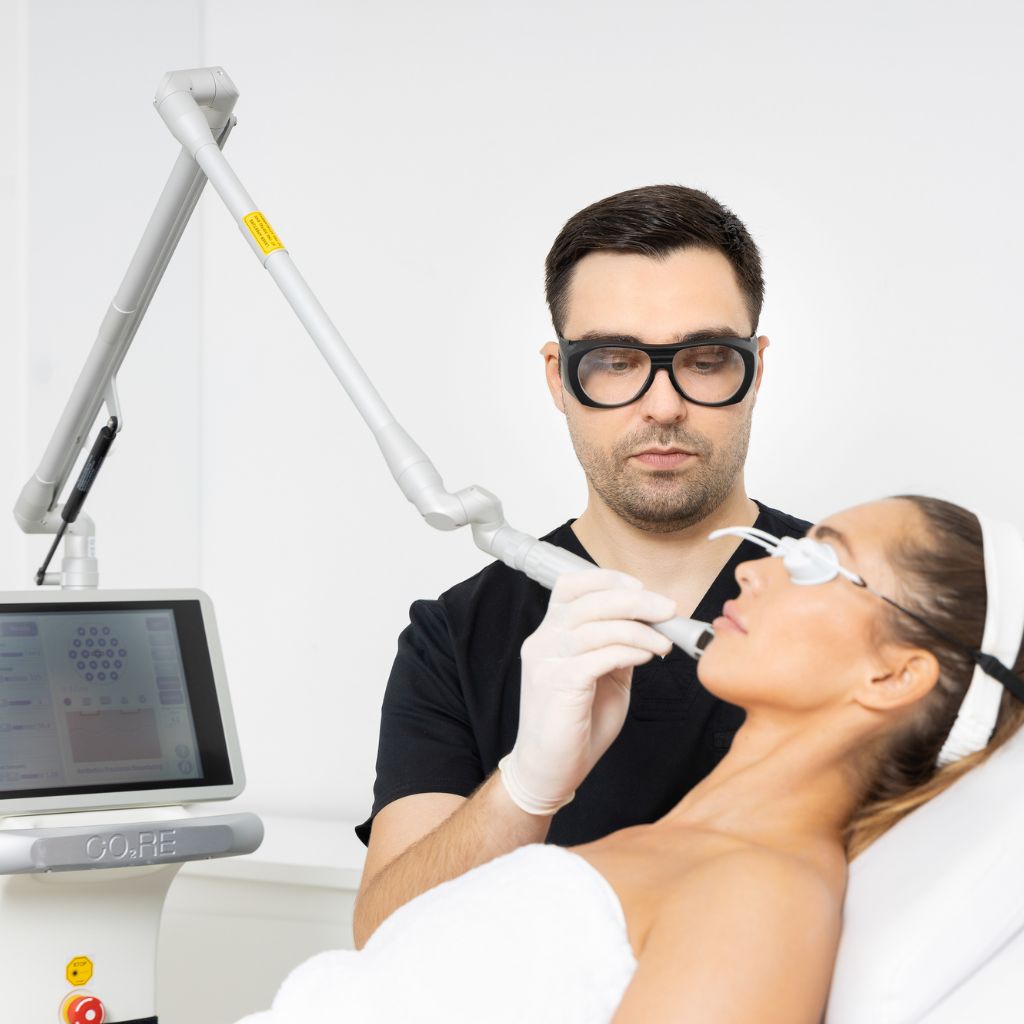
If the scar remains more pronounced or uneven, it can be treated with a CO2 laser; the dermatologist decides on this once the scar has matured.
Radiofrequency mole removal price
Current prices are listed in our dermatology price list. The price depends on the number, size and location of the lesions, and the removal plan is determined after an examination.
Our dermatologists
Who performs radiofrequency mole removal?
Have a question?
Frequently asked questions about radiofrequency mole removal
Is the radiofrequency method suitable for every mole?
No. Suitability depends on the type, depth, size and location of the lesion. A dermatologist assesses this during the consultation and, if the radiofrequency method is not the best choice, suggests an alternative procedure.
Does radiofrequency mole removal hurt?
Pain is not felt during the procedure, as an anaesthetic cream is applied beforehand or a local anaesthetic is administered. The injection when administering the anaesthetic can be unpleasant, and the area may be temporarily sensitive after the procedure.
When can I wear make-up or shave over the treated area?
Only when the wound has completely healed and the scab falls off on its own, usually after 7 to 10 days. Until then, that area must not be shaved, and make-up must not be applied directly to the wound.
When can I exercise, swim or expose the area to the sun?
Workouts that cause heavy sweating, swimming pools and saunas should be avoided until the wound has healed. The treated area should be protected from the sun for a few months in order to reduce the risk of skin darkening.
Can a mole reappear?
Yes, because the radiofrequency method, unlike the surgical one, does not guarantee the removal of all mole cells in the deeper layers of the skin. If pigment reappears in the same place, the lesion should be shown to a dermatologist, along with the report from the previous histopathological analysis, in order to decide on the further course of action.
When should I contact a doctor after the procedure?
If the wound bleeds and the bleeding does not stop after applying pressure, if increased redness, swelling, warmth or pus appear, if you have a high temperature, or if the wound does not heal within the expected timeframe.
Can multiple moles be removed in a single appointment?
Yes, when the doctor assesses that it is safe. The number of lesions depends on their size, location and the overall scope of the procedure.
When is the procedure postponed?
The procedure is postponed in the event of a skin infection or inflammation at the site of removal, and in cases of acute illness. If a pacemaker, defibrillator or other electronic medical device is fitted, a special assessment is required, as this depends on the type of device and the way radiofrequency energy is applied. Chronic conditions, such as diabetes or epilepsy, are evaluated individually by the physician. During pregnancy, removal for cosmetic reasons is postponed until after pregnancy.
Book radiofrequency mole removal in Belgrade
Imedic poliklinika is located at Kneza Miloša 23, 2nd floor, Belgrade. Opening hours: Monday–Sunday, 9:00–21:00.
Appointments can be booked by phone: 064 237 0707. If you contact us from abroad, we are also available via Viber and WhatsApp on the same number.

Author: Dr Ivica Jeremić
PhD in Medical Sciences (Molecular Medicine and Immunology), rheumatology subspecialist, dermatology resident.
Updated:
Expert sources
- Curley RK, Cook MG, Fallowfield ME, Marsden RA. Accuracy in clinically evaluating pigmented lesions. BMJ. 1989;299(6690):16–18.
- Use of antibiotic-containing ointment versus plain petrolatum during and after clean cutaneous surgery. Journal of the American Academy of Dermatology (PubMed 17052511).
- Management of cardiac implantable electronic devices around the time of dermatological procedures requiring electrosurgery: an important guidance update (PubMed 38038380).
Professional dermatological supervision and reliable information
The text on this page was written in accordance with the clinical practice of Imedic poliklinika and verified by a specialist in dermatovenereology. The expert sources are listed above. The doctor decides on removing a mole after a clinical and dermoscopic examination, along with an assessment of the type of lesion and its behaviour over time. In case of any doubt, surgical removal with histopathological analysis is preferred. The procedure plan, wound care and follow-ups are determined individually. An in-person examination by a dermatologist is essential for the assessment.