Surgical mole removal in Belgrade
Surgical removal of a mole is a procedure whereby the lesion is removed in its entirety and sent for histopathological analysis. Following a clinical and dermoscopic examination, the dermatologist assesses whether removal is necessary and which approach is appropriate for the specific lesion. The intervention is performed under local anaesthesia, and after the procedure the patient receives instructions for wound care, follow-up and removal of stitches.
Surgical mole removal at a glance
| When it is done | When the examination findings require the lesion to be analysed or when a harmless mole is bothersome |
|---|---|
| Anaesthesia | Local |
| Duration of the procedure | About 60 minutes, depending on the size, location and number of lesions |
| Method | Elliptical excision with a margin of healthy tissue and primary closure with sutures |
| Analysis | Removed tissue is always sent for histopathological analysis |
| Procedure performed by | Dermatovenereology specialist |
What are moles?
Moles (naevi) are most commonly harmless skin lesions caused by the accumulation of melanocytes – the cells that produce the pigment melanin. Most adults have between 10 and 50 moles, and only a small number of them require further diagnostics or removal. More information on the formation of moles, monitoring them and the reasons for removal can be found in our article Everything you need to know about moles.
When is surgical removal of a mole recommended?
The decision on removal is made after an examination, never in advance. The first step is dermatological examination, during which the lesion is assessed and further steps are planned; before any decision on removal, the doctor performs a dermoscopic mole check.
Medical reasons
Surgical removal is recommended when a dermoscopic finding reveals a lesion that needs to be analysed: an irregular or asymmetrical mole, multiple colours within the same lesion, a mole that is growing or changing, bleeding, or failing to heal. In such cases, removal is a diagnostic step, as only tissue analysis provides a definitive answer regarding the nature of the lesion.
Aesthetic and practical reasons
A harmless mole can also be removed when it is bothersome – it is located in an area of friction, catches on clothing or jewellery, gets injured during shaving, or the patient simply does not like its appearance.
Surgery or another method?
The surgical method enables the lesion to be removed in its entirety and sent for analysis, making it the method of choice for any lesion that needs to be examined. For certain lesions assessed as benign, the doctor may consider mole removal by radiofrequency. Tissue-destroying methods, such as laser vaporisation, leave no sample for analysis, so they are not used when the lesion needs to be examined. Surgery is also used to remove atheromas, which are commonly referred to as sebaceous cysts.
Examination and pre-procedure preparation
Before the procedure, the dermatologist examines the lesion, explains the procedure and gives instructions for preparation. It is important to tell the doctor:
- which medicines and supplements you take, especially blood thinners;
- whether you are allergic to local anaesthetics, disinfectants or plasters;
- whether you bruise or bleed easily;
- whether you have previously had problems with wound healing or prominent (keloid) scars;
- whether you are pregnant or have any chronic illnesses.
Bring any previous medical reports and test results, if you have them. Do not stop prescribed medication on your own; any changes will be decided by the doctor.
What does surgical mole removal involve?
1. Examination and procedure plan
A dermatologist examines the lesion with a dermatoscope, explains the reason for removal, and plans the size and direction of the incision so that the scar is as inconspicuous as possible.
2. Local anaesthesia
The site of the procedure is disinfected and a local anaesthetic is administered. Only the needle prick of the anaesthetic injection is unpleasant; during the removal itself, no pain is felt.
3. Removing the lesion
The mole is removed by elliptical excision, entirely and with a narrow margin of healthy tissue, so that the analysis can confirm that the lesion has been completely removed.
4. Wound closure
The wound is closed with stitches and covered with a waterproof sterile dressing.
5. Histopathological analysis
The removed tissue is sent to the laboratory for histopathological analysis, which provides definitive information on the nature of the lesion.
6. Instructions and follow-up
The patient receives wound care instructions and appointments for dressing changes, suture removal and a follow-up consultation at which the dermatologist will explain the findings.
Why is a histopathological analysis performed?
Histopathological analysis is the examination of removed tissue under a microscope. The pathologist determines the nature of the lesion, whether it is benign, and whether it has been completely removed with a sufficient margin of healthy tissue. This is the only method that provides a definitive diagnosis, which is why every surgically removed lesion is sent for analysis – even when it has been removed for aesthetic reasons.
Surgical removal with tissue analysis is also an excisional biopsy; more information on sample collection methods can be found on our page about skin biopsy.
The test result is usually available within a few working days to two weeks, depending on the laboratory and the type of lesion. The result is interpreted by a dermatologist, who explains at a follow-up appointment what has been found and whether further steps are required.
Recovery, wound care and removal of stitches
Following the procedure, a few stitches and a waterproof sterile dressing remain at the removal site. Recovery depends on the location and extent of the procedure, and usually looks like this:
- the first two days – the dressing is not removed and the wound must not get wet;
- first dressing change – the dressing is changed and from then on the wound can be washed, according to the doctor's instructions;
- until the stitches are removed – avoid physical exercise, swimming pools and saunas;
- removal of stitches – after 7 days on the face, and after 14 days on other parts of the body.
Most patients return to activities that do not involve physical exertion the same or the following day. A doctor should be contacted if the wound bleeds and the bleeding does not stop after applying pressure, if increased redness, swelling, warmth or pus appear, if there is a raised temperature, or if the wound starts to come apart.
Will it leave a scar?
Following surgical removal, a thin, linear scar remains. Its final appearance depends on the site and size of the procedure, skin tension in that part of the body and individual healing – on the face, the scar is usually less noticeable than on the back or chest.
The scar matures gradually, over months: initially it is pink, and over time it fades. Sun protection during this period helps to prevent it from darkening. Removal without a scar cannot be promised, but the incision is planned so that the scar is as inconspicuous as possible.
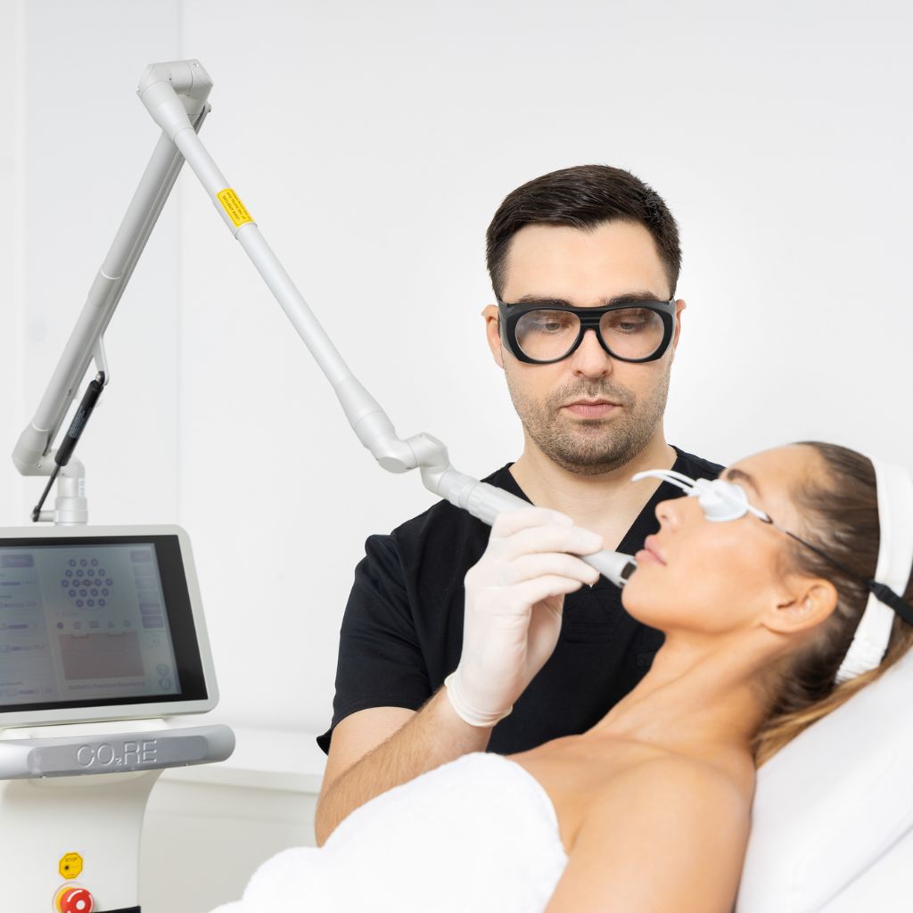
If the scar remains prominent, raised or uneven after maturing, it can be treated with a CO2 laser, which stimulates skin renewal and evens out its surface. A dermatologist determines when the right time is for the treatment and how many treatments are needed.
Cost of surgical mole removal
Current prices are listed in our dermatology price list. The price depends on the number, size and location of the lesions, and the intervention plan is determined after an examination.
Our dermatologists
Who performs surgical mole removal?
Have a question?
Frequently asked questions about the surgical removal of moles
Does the surgical removal of a mole hurt?
The procedure is performed under local anaesthesia, so no pain is felt during the removal. Generally, only the injection when administering the anaesthetic is unpleasant. Once the effects of the anaesthesia wear off, the site of the procedure may be sensitive for a few days.
Is the procedure safe?
Surgical removal of a mole is a routine procedure under local anaesthesia and complications are rare. Bleeding, infection, wound reopening or a more prominent scar are possible, which is why it is important to follow the wound care instructions and attend the follow-up appointment.
Can multiple moles be removed in a single appointment?
Yes, if the doctor considers it safe. The number of lesions removed in a single appointment depends on their size, location, and the overall scope of the procedure.
How is a mole on the face removed?
On the face, the incision is planned along the lines of natural skin tension so that the scar is as inconspicuous as possible, and the stitches are removed earlier, after 7 days. For facial moles assessed as harmless, the doctor may also consider radiofrequency removal.
When can I return to exercise?
Physical exercise, the swimming pool and the sauna should be avoided until the stitches are removed, as strain and moisture put stress on the wound. After the stitches are removed, activities are gradually reintroduced, following the doctor's advice.
Can a mole reappear?
When the lesion is removed in its entirety, with a margin of healthy tissue, recurrence is rare. If pigment or a new lesion appears at the site of the scar, it should be shown to a dermatologist.
What happens when the test result arrives?
The histopathological analysis report is interpreted by a dermatologist, who explains what has been found and whether further steps are needed. In most cases, the report is normal and no further measures are necessary. If the findings require it, the doctor recommends re-excision or further monitoring.
When is the procedure postponed?
The procedure is postponed in the event of a skin infection or inflammation at the removal site and in cases of acute illnesses. Treatment with blood-thinning medication is not an automatic obstacle – the plan depends on the type of treatment, the scope of the procedure, and individual risk. Do not stop prescribed medication on your own; any changes are decided by the doctor.
During pregnancy, surgical removal is performed when the dermoscopic finding indicates the need to analyse the lesion, whereas removal for aesthetic reasons is postponed until after pregnancy.
Book a mole check and removal in Belgrade
Imedic poliklinika is located at Kneza Miloša 23, 2nd floor, Belgrade. Opening hours: Monday–Sunday, 9:00–21:00.
Appointments can be booked by phone: 064 237 0707. If you contact us from abroad, we are also available via Viber and WhatsApp on the same number.

Author: Dr Ivica Jeremić
PhD in Medical Sciences (Molecular Medicine and Immunology), rheumatology subspecialist, dermatology resident.
Updated:
Expert sources
- Curley RK, Cook MG, Fallowfield ME, Marsden RA. Accuracy in clinically evaluating pigmented lesions. BMJ. 1989;299(6690):16–18.
- Lewis KG, Dufresne RG Jr. A meta-analysis of complications attributed to anticoagulation among patients following cutaneous surgery. Dermatologic Surgery. 2008;34(2):160–164.
- Sequeira Campos MB, Xiao A, Ettefagh L. Laser Revision of Scars. StatPearls. Treasure Island (FL): StatPearls Publishing; 2025.
Professional dermatological supervision and reliable information
The text on this page is written in accordance with the clinical practice of Imedic poliklinika and verified by a specialist in dermatovenereology. The expert sources are listed above.
The decision to surgically remove a mole is made by a doctor after a clinical and dermoscopic examination. Every removed lesion is sent for histopathological analysis, which is the only way to provide a definitive diagnosis.
The treatment plan, wound care, and follow-up are determined individually, based on the findings and the patient's state of health. An in-person examination by a dermatologist is required for an assessment.