Moles are one of the most common skin changes and almost everyone has them, but how much do we actually know about them? Dr Marina Ćirović, a specialist in dermatology and venereology at our clinic, explains what moles are, why their examination is important and when they should be removed. Moles are benign structures made up of cells called melanocytes. These are cells ...

Moles are one of the most common skin lesions and almost everyone has them, but how much do we actually know about them? Dr Marina Ćirović, a dermatovenereology specialist at Imedic poliklinika, explains what moles are, why their examination is important, and when they should be removed.
Moles are benign structures made up of cells called melanocytes. These are cells present in our skin, as well as in some other tissues — such as the membranes of the eye and the meninges. When located in the skin, melanocytes are responsible for producing the pigment melanin, which gives our skin its colour and complexion.
What determines how a mole looks is the position of the melanocytes in the skin. When they are organised superficially, in the so-called basal layer of the epidermis, they form moles that are flat, level with the skin — most commonly recognised as round or oval spots of various shades of brown. When melanocytes are organised deeper, in the dermis, raised moles are formed that partially sit above the skin level. It is these raised moles that often confuse patients — they can resemble fibromas or warts, so many people come for an examination thinking it is some other skin lesion.
When an examination determines that it is a viral wart, the method of treatment is chosen according to its type and location. More about this can be found on our page about wart removal.
Why is a mole check important — and what is a dermatoscope?
Moles on the skin can resemble other lesions, and other lesions can resemble moles. Precisely for this reason, examinations are not performed with the naked eye, but with the aid of a device called a dermoscope or dermatoscope. Essentially, it is a magnifying glass that uses different levels of magnification and — crucially — polarised and non-polarised light. This enables the dermatologist to see structures that are not visible to the naked eye and thereby distinguish moles from all other skin lesions.
And why is that distinction so important? Because this spectrum of lesions includes both benign and malignant ones. Malignant lesions — skin cancers and particularly melanoma, which arises precisely from melanocytes — need to be removed so that they do not threaten general health. Melanoma is a very aggressive malignant tumour, but if detected in time, its removal saves lives.
Therefore, it is recommended that everyone over the age of 20 should have a preventive full-body skin examination once a year — and not just a visual examination with the naked eye, but a dermoscopic examination. It serves as a primary screening for the early detection of melanoma.
Digital dermoscopy — monitoring moles over time
There are situations when a dermatologist cannot immediately make a final decision based on a single examination — especially when it comes to pigmented lesions, very small lesions, or normal moles that show signs of growth. In such cases, it is not enough to look at the lesion just once; it needs to be monitored over time. This means carrying out an examination today, and then reviewing that same lesion in a month, in three months, or in six months — and determining whether, and in what way, it is changing.
To ensure this monitoring is accurate, the lesions need to be photographed. Photographs can be taken with a smartphone too, but such a storage system is impractical, and the image quality is generally poorer. This is precisely where the digital dermatoscope comes in. Digital dermoscopy allows the monitoring of moles and other skin lesions in high resolution and at a much higher magnification than conventional hand-held dermoscopy provides. The photographs are stored in software designed exclusively for this purpose, which allows for greater precision in diagnosis. At Imedic poliklinika, we use the Canfield D2 dermatoscope, providing exceptionally high image quality at different levels of magnification and with different light polarisation.
When a follow-up image is taken after three or six months and compared with the previous one, it is possible to spot even the most subtle changes — those that we would never see with the naked eye. If it is established that the lesion is changing, a decision is made to remove it and send it to a pathologist for examination.
It is important to emphasise that it is not necessary to photograph all moles. Only those suspected of changing are photographed. The very purpose of digital dermoscopy is to reduce the number of unnecessary excisions. For individuals who do not want scars or unnecessary interventions, monitoring changes over time is a way to intervene only when it is truly necessary.
More about capturing and saving photographs and comparing lesions at follow-up visits can be found on our page about digital dermoscopy.
Who is at increased risk of melanoma?
Based on the initial dermoscopic examination, the dermatologist assesses how many moles the patient has, what they are like, and whether there are any atypical moles. Based on this, the risk category is determined — for both the patient and the doctor — and a decision is made regarding how often follow-up visits should take place and whether it is necessary to photograph a larger number of lesions.
For people who are not at increased risk, a preventive check-up once a year is quite sufficient. For those at increased risk, more frequent follow-up visits and more detailed monitoring are recommended.
So who falls into that increased risk group? Dr Marina Ćirović highlights several factors:
- Fair skin and skin phototype — individuals who easily burn in the sun but almost never tan, who have light eyes and fair hair, are classified as skin phototypes I and II and carry a higher risk.
- A large number of moles — having more than 50 moles is considered to increase the risk of developing melanoma.
- Family history — individuals who have a close relative in the family who has had melanoma, or have themselves had melanoma earlier in life, are at increased risk.
- Previous malignant skin tumours — people who have had other types of skin cancer are also in the high-risk group.
- Childhood sunburn — sun exposure and sunburns before the age of 18 are considered a factor that increases the risk of later developing melanoma.
- The use of sunbeds — artificial UV radiation, just like natural sunlight, increases the risk of melanoma, regardless of skin phototype.
How are moles removed?
When the decision is made that a certain mole needs to be removed, the removal method depends on the type of mole it is and the result of the dermoscopic examination.
When there is a suspicion of malignancy or in the case of an atypical mole, removal is always performed surgically — with a scalpel. Surgical removal entails excising the mole along with a small margin of surrounding healthy skin, and closing the wound with stitches, which are removed after about two weeks. The wound is left dry for the first two days, and at the first dressing change, the doctor assesses whether the patient can continue wound care at home or should attend dressing changes until the stitches are removed. The tissue that is removed is always sent to a pathologist for examination — and this is a rule without exception.
More detailed information regarding the procedure, histopathological analysis and recovery can be found on our page about surgical removal of a mole.
When a dermoscopic examination is completely normal and a mole is removed at the patient's request — most frequently for aesthetic reasons or because it is bothersome and catches on things — in certain cases, it is possible to apply radiofrequency removal. This method is particularly suitable for smaller pedunculated (hanging) moles whose base allows it.
At the moment of tissue separation, the radiofrequency knife coagulates blood vessels, which means the wound does not bleed and does not require stitches. Wounds from radiofrequency removal heal spontaneously, most often within two weeks, depending on the size of the lesion. The patient is given wound care instructions to follow at home.
Even in the case of radiofrequency removal, the tissue is sent to a pathologist — a small margin of healthy surrounding skin is always removed as well so that the mole tissue reaches the pathologist intact.
It is important to emphasise that radiofrequency removal is used exclusively for moles that are dermoscopically completely normal, without any signs of change or activity. Everything else is removed surgically.
Find out more — watch the video by Dr Marina Ćirović
Moles are an integral part of our skin and most of them are completely harmless. However, as Dr Marina Ćirović explained, a regular dermoscopic examination is not something that should be put off — because with the naked eye it is simply not possible to see what a dermatoscope sees.
Once a year, a preventive full-body skin examination is sufficient for most people.
For those at increased risk, a dermatologist will recommend more frequent check-ups and digital monitoring of changes over time. The aim is not to remove as many moles as possible — the aim is to identify in good time those that genuinely require intervention.
If you haven't had a check-up for a while, or notice that a mole is changing — don't wait. Book a consultation at Imedic poliklinika and have peace of mind about your skin's health.
Everything Dr Marina talked about can also be watched in the video below.
IMEDIC NOVOSTI
Be among the first to know about discounts and special offers
Sign up and receive notifications about current promotions, new treatments and useful advice from our doctors.
DISCOUNTS · NEW OFFERS · DOCTOR'S ADVICE
Sign-in is currently unavailable. Please try again in a few moments.
One more step: check your email and click on the confirmation link.
Want to see our new texts on Google more often?
Add imedic.rs as a preferred source in Google Search. When we publish a new text on skincare, aesthetic medicine or dermatology, Google will show it to you more frequently among the top stories. Setup takes a few seconds and can be changed at any time.
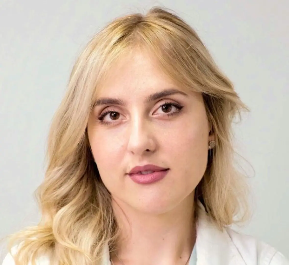
Dr Marina Ćirović
The Imedic team writes educational articles on dermatology, aesthetic medicine and skincare – clear, reliable information for patients.
Updated: