Has a mole changed its colour, shape or size? Find out when to book an appointment, what to record and how photographs can help the dermatologist.

If a mole you have known for years has changed colour, shape, size or texture, book an appointment with a dermatologist. The same applies if it has started to itch, bleed or ooze. When booking, state that you are coming because of a change in an existing mole. [2, 3, 4]
A change does not automatically mean that it is a melanoma, a malignant skin tumour. Benign moles can also change over the course of a lifetime, but the cause of a specific change is assessed through an examination. You do not need to name the problem yourself — it is enough to describe what you have noticed. [1, 5]
What changes in a mole should be shown to a dermatologist?
Describe to the dermatologist what has changed compared to the previous appearance of the mole. The change may affect the entire mole or just one part of it. A table can help you describe it; the examples given are not diagnoses. [2, 3]
| What have you noticed? | How can you describe change |
|---|---|
| Colour | The mole has darkened, lightened or a new shade has appeared in one part. |
| Shape and edges | One side looks different, the outline has changed or the edge has become irregular. |
| Size or new emergence | The mole has enlarged, is growing in one direction or a new raised part has appeared. |
| Area | The skin over the mole is flaking, a small crust or sore is appearing, and the mole is weeping or bleeding. |
| New symptoms | Itchiness, sensitivity or pain has appeared that was not previously present. |
There is no need to wait for more signs to appear. Even a single new change is sufficient reason to consult a doctor. [1]
Should one wait for the routine check-up?
Report any new changes before your already scheduled routine check-up. A planned appointment in a few months does not mean that every new observation should be left for that visit. If you were recently examined, please also state when it took place. [4, 6]
When booking, specifically emphasise rapid growth, bleeding or a sore that does not heal. [5, 13] State how long it lasts, whether it is getting worse, and whether the bleeding was preceded by an injury. That description helps the medical staff determine the priority of the examination. [4]
Monitoring is planned after an examination, when the dermatologist considers it to be the appropriate next step. At that point, the findings for the chosen lesion are compared over time, alongside a booked follow-up appointment. If you notice a new lesion during the monitoring period, please contact us sooner; the doctor will assess whether the appointment needs to be brought forward. [6, 7]
What to note down before the check-up?
Prepare a short note on the course of the change and the documentation you already have. You do not need to know the exact date or measure every detail; it is helpful to separate what you remember reliably from what you are unsure of. [6]
- When did you first notice the change: state the approximate date or period and who spotted it.
- What has changed: describe what made you pay attention.
- How it developed: has it remained the same since then, become more pronounced or, for example, does the itching occasionally return.
- Was there any injury or friction: mention shaving, scratching, catching on clothing or any other event that preceded the change. [5]
- Are there any earlier findings and photographs: have them ready or prepared on your phone, along with the date if known. [4, 10]
For example, you could say: „I noticed the darker patch last week, but I don't know when it appeared.” By doing so, you clearly distinguish the moment of noticing from the possible start of the change. You don't have to fill in the gaps in your memory.
Tell the doctor even if you have previously had melanoma or another skin cancer, or if a close family member has had it. This is information that complements the discussion about the specific mole. [4, 8]
Can a photograph of a mole help?
A photograph can help to document appearance, but its value depends on the time it was taken and the quality of the image. If you do not have an old photograph, the review can be carried out without it. [6, 9]
Older photograph you can show what the mole looked like previously, provided it can be seen clearly enough. Keep the original and the date if you know it; do not change the colours or edit that part of the image.
New home photography Record the present appearance. Use good lighting, focus the image and turn off filters. One wider shot showing the location on the body and one closer-up shot are useful. Shadow or glare can obscure details, so check that what you noticed on the skin is clearly visible in the footage. Photographing should not delay a necessary appointment. [10]
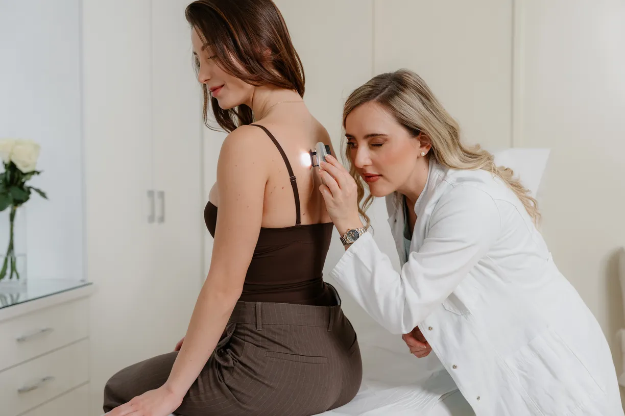
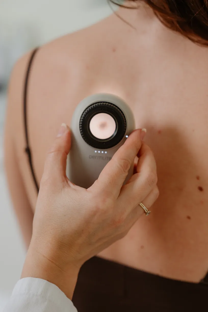
Dermoscopic photograph the doctor examines using a skin magnification device. Within digital dermoscopy, Selected images are kept for comparison during check-ups. A regular phone photograph does not show the same level of detail and is not a substitute for an examination or a basis for ruling out a serious condition on your own. [7, 9, 14]
How does a dermatologist assess a mole that has changed?
The dermatologist assesses the current appearance of the mole and information on how it has changed. During the conversation with you, they clarify details regarding the course of the change, previous findings, your state of health, and the medication you are using. They then examine the mole and the surrounding skin, and, if necessary, other lesions on the body. [8, 11]
During dermoscopic examination of moles a dermatoscope is used, a device that magnifies and illuminates the lesion so that the doctor can see details that are harder to spot with the naked eye. Examination with this device does not involve tissue removal. Available earlier images and findings can complement the assessment. [7, 9]
Based on the overall findings, the dermatologist recommends the next step: advice and follow-up, targeted monitoring, or removal for analysis. Dermoscopy helps with this decision, but does not replace tissue examination when there is a medical reason for it. [7, 11]
Before leaving, clarify what has been agreed, when the next step is, and who to contact if you have a question about the test result. If there is something you have not understood, ask the doctor to explain the recommendation more simply or write it down. [4, 6]
Must a changed mole be removed?
Not every changed mole needs to be removed. If a dermatologist recommends removal to clarify the diagnosis, the removed tissue is sent for histopathological analysis — examination of a tissue sample under a microscope by a pathologist. [7, 11]
Details of the procedure are explained on the page surgical removal of a mole. Agree on how you will get the result and who will interpret it for you, as further recommendations depend on the findings. [12]
Have a question?
Answers to frequently asked questions
Is a mole less concerning if it does not hurt?
The absence of pain is not a reliable indicator of the nature of a lesion. Melanoma can exist without pain or discomfort, so the physical sensation on the skin cannot replace the assessment of the appearance and development of a mole. [13]
Is it important that the mole is very small?
Size is one of the data points, but a small diameter by itself does not confirm that a lesion is harmless. Melanoma can be smaller than six milimetres. Therefore, that number should not be used as a threshold below which an examination is unnecessary. [1]
For booking mole checks at IMedic contact the practice and state that you would like an assessment of a mole that has changed. Information on how to book is available on the examination page.
Expert sources
- American Academy of Dermatology. Moles: Signs and symptoms.
- National Cancer Institute. Common Moles, Dysplastic Naevi, and Risk of Melanoma.
- NHS. Moles.
- American Academy of Dermatology. How to choose a dermatologist.
- DermNet. ABCDEFG of melanoma.
- Cancer Research UK. Seeing your GP for symptoms of melanoma skin cancer.
- Nice. Melanoma: assessment and management — NG14.
- NHS. Symptoms of melanoma skin cancer.
- Cancer Research UK. Tests for melanoma skin cancer.
- American Academy of Dermatology. How to take pictures of your skin for your dermatologist.
- American Academy of Dermatology. Skin cancer types: Melanoma — Diagnosis and treatment.
- NHS. Tests and next steps for melanoma skin cancer.
- American Academy of Dermatology. Skin cancer types: Melanoma — Signs and symptoms.
- NHS England. A teledermatology roadmap: implementing safe and effective teledermatology triage pathways and processes.
IMEDIC NOVOSTI
Be among the first to know about discounts and special offers
Sign up and receive notifications about current promotions, new treatments and useful advice from our doctors.
DISCOUNTS · NEW OFFERS · DOCTOR'S ADVICE
Sign-in is currently unavailable. Please try again in a few moments.
One more step: check your email and click on the confirmation link.
Want to see our new texts on Google more often?
Add imedic.rs as a preferred source in Google Search. When we publish a new text on skincare, aesthetic medicine or dermatology, Google will show it to you more frequently among the top stories. Setup takes a few seconds and can be changed at any time.

Imedic
The Imedic team writes educational articles on dermatology, aesthetic medicine and skincare – clear, reliable information for patients.
Updated: